First area: Intermediate positioning splints
We make intermediate positioning trays for bimaxillary surgery, according to the decisions of the surgeon who requests them.
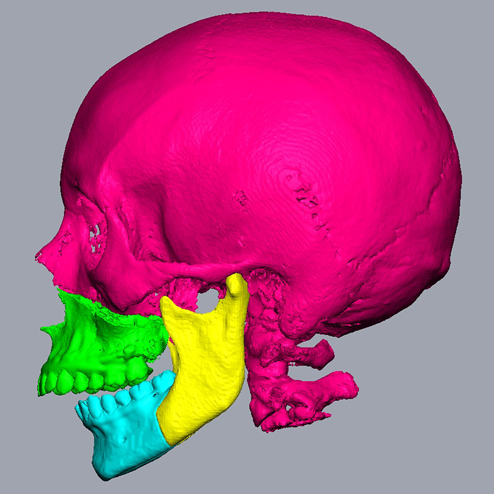
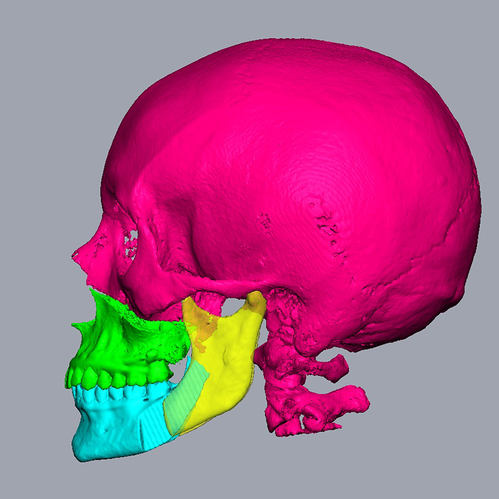
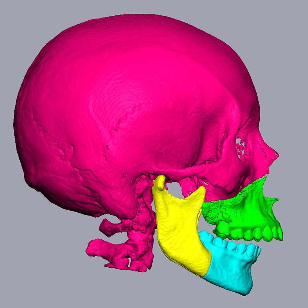
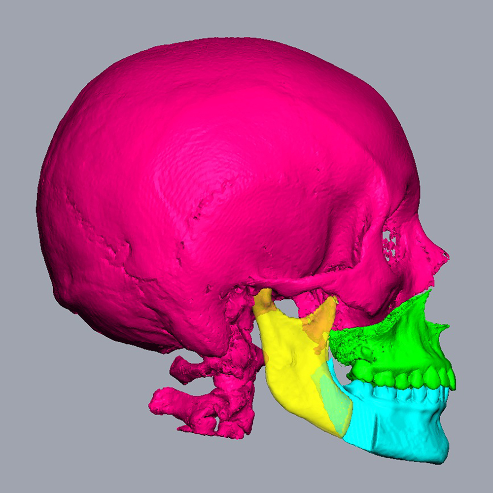
Working closely with your team, we design these trays based on the recommendations provided. Our process uses DICOM files from the facial scanner, which we link to STL files of digital impressions of the dental arches. If you have any doubts or uncertainties about the quantification of displacements, we offer the option of providing several simulations.
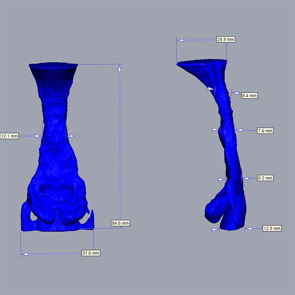
We provide the various stages of the process with 3D digital images from different angles: left profile, right profile, occlusal view, as well as intermediate positions after mobilisation of the maxilla (maxillary primary surgery) or mandible (mandibular primary surgery). We also offer quantitative analyses detailing maxillary and mandibular movements in all three spatial dimensions, enabling you to make informed decisions.
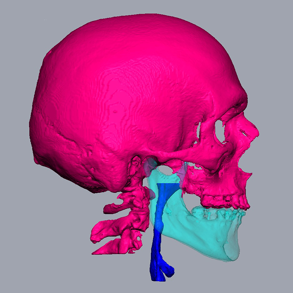
We can also provide you with a life-size 3D print of your patient’s skull.
Our treatment proposal is not binding on 3D Vision Ortho. It must be validated by the surgeon in charge of the patient, who is in fact solely responsible.
A quotation will be sent to you based on your request and the number of patients for whom you require expertise.